By Dr. Chris Cebra, Oregon State University
I am writing this editorial to describe Eimeria
macusaniensis infection, which I believe to be one of the most important emerging diseases in camelids. The
causative organism is the largest of the camelid coccidia, 100 microns in length, thick-walled, and
watermelon seed-shaped. It has longer prepatent (32 to 43 days) and patent (up to 40 days) periods than other
species of coccidia, and also is as commonly associated with disease and death in adults as in
crias. It is not a new organism. Dr. Jarvinen's work from 1999 suggested that it was present in most herds,
and my recent experiences support that. It is "emerging" because of greater awareness, better
detection methods, and possibly higher case attack rates. It is my belief that the extreme environmental
stability of this organism has allowed it to build up over the 20+ years of popular camelid farming in the U.S., and that together with certain management
practices including overcrowding, co-housing of crias and adults, overuse of certain pastures, and mixing
stressed camelids from different locations, has lead to a real increase in clinical cases.
Eimeria macusaniensis follows the typical coccidian lifecycle, requiring 13 to 21 days on the ground for
oocysts to sporulate, having multiple rounds of replication within the host, and primarily damaging
the intestinal epithelium. The severity of clinical disease relates to host immunity and infective dose.
Lightly infected, immunocompetent camelids may transiently shed small numbers of organisms, but will
show no signs. Immunocompromised or immunonaive camelids, such as stressed adults or all
crias, confronted with a large or overwhelming dose, may succumb to fatal disease in as little as 3 weeks, or
as long as 2 weeks before oocysts appear in the feces. Adults within an infected herd appear to be less
susceptible than ones introduced later, for example for breeding. The major signs of severe disease are
those of weakness and wasting. Unless the camelid is weighed frequently, the owner frequently notices
nothing wrong until the camelid collapses. Diarrhea is uncommon, except in crias. Hypoproteinemia is the
major blood abnormality. Clinical abnormalities will be more comprehensively described in a
soon-to-appear scientific report.
The combination of lack of specific GI signs and a negative fecal examination makes the infection easy to
miss. We consider all ill-thrift camelids to be suspect, and either treat empirically or perform
multiple fecal examinations over at least a 2 week period. Even then, shedding is often light (<100
oocysts/g) for the first week of patency, even in camelids with overwhelming infections. Considering
that most of the anticoccidial medications available in the U.S. are most efficacious against the earlier
stages of the organism, delaying treatment in suspect camelids is unlikely to be advantageous. It is more
difficult to decide what to do with non-clinical shedders and their herdmates. As with other internal
parasites, in general we acknowledge their presence and try to control them, not eliminate them. The long
prepatent period, together with the pelleted camelid feces, offers the potential for timely manure removal
to decrease pasture contamination. We believe the thick wall of the organisms imparts extreme
environmental resistance, so leaving paddocks and pastures empty is of less benefit than with other GI
parasites. Radical soil treatments including burning and topsoil removal have been tried, but such
approaches should only be considered in extreme circumstances. As with other parasites, a good
management approach decreasing stocking density, avoiding admixtures of stressed camelids, encouraging
eating from above-ground feeders, avoiding excess cohabitation of crias and adults, conducting fecal
parasite surveillance, timely antiparasitical treatments, and frequently checking demeanor and body condition of camelids should decrease the number of affected camelids and allow early identification of
those that require treatment.
I have only lightly addressed fecal detection. This will also be the subject of an upcoming scientific
report. The high density of the oocyst makes it more likely to float in denser float solutions and also
slower to rise. The low numbers at the onset of patency dictate that very sensitive techniques should
be used. These factors should be considered when choosing the method of fecal analysis.
Primary Reference:
Cebra CK, Valentine BA, Schlipf Jr. JW, Bildfell RJ,
McKenzie E, Waitt LH, Heidel JR, Cooper BJ, Lohr CV,
Bird KE, Saulez MN, Firshman AM. "Eimeria
macusaniensis infection in 15 llamas and 34 alpacas".
J Am Vet Med Assoc. 2007 Jan 1;230(1):94-100.
Reprinted from Wool & Wattles: The AASRP Newsletter,
vol. 34, #4; pp. 10-11, Oct.-Dec. 2006 with
permission from Dr. Chris Cebra and the American
Assoc. of Small Ruminant Practitioners. Permission to
reprint this article is granted to participants in the
Newsletter Exchange.
~~~~~~~~~~~~~~~~~~~~~~~~~~~~~~~~~~~~~~~~~~~~~~~~~~~~~~~~~~~~~~~~~~~~~~~~~~~~
From Camelid Healthcare Services:
Eimeria Macusaniensis (E.mac) is a coccidian parasite that can
cause severe illness. E. mac is called a �large coccidia� because
its oocyst (egg) is very large compared to the other coccidia of
llamas and alpacas. E. mac, like the other �small� coccidia is a
protozoan parasite that lives in the intestinal tract. This
parasite causes damage to the lining of the intestine and inhibits
normal absorption of nutrients and causes fluid to be secreted
from the damaged intestinal wall. This results in diarrhea, weight
loss and low blood protein levels.
E. mac is a common parasite of many alpacas and most farms will
have some positive animals. A low number of E. mac found on a
routine fecal exam is not cause for alarm�but something that
should be noted. Most researchers would agree that a low level
exposure to parasites results in some protective immunity and is
desirable. Health problems occur when an animal is exposed to an
overwhelming load of parasites or if exposure occurs when the
animal�s natural defenses are low. This may occur if a cria failed
to get a good passive transfer of colostrum, during show season or
with travel to a new farm.
Diagnosis of illness caused by E. mac can be difficult because of
the long pre-patent period. This pre-patent period is the time
from infection (and damage to the intestinal wall) till the time
the oocysts appear in the feces. Finding the oocysts in the fecal
exam and clinical disease are the criteria that we use to
establish the diagnosis. E. mac will have a pre-patent period of
35-43 days�so the animal can have damage to the intestine and we
can not prove it is caused by E. mac because the fecal exam is
negative.
Many times treatment for E. mac is started based on farm history
of E.mac being present, clinical signs of diarrhea and low blood
proteins on examination of blood work.
Treatment of E. mac can be accomplished with Corid, sulfa drugs or
a new product called Marquis. Consult with your veterinarian for
best treatment for your particular situation.
~~~~~~~~~~~~~~~~~~~~~~~~~~~~~~~~~~~~~~~~~~~~~~~~~~~~~~~~~~~~~~~~~~~~~~~~~~~~
By Jill McElderry-Maxwell, Bag End
Suri Alpacas of ME, LLC (2010)
(Editor�s Note: The author explains that she is not
a veterinarian, and her intent with the article was to pull
together the research available on the topic so that individuals
could make informed decisions about their parasite management
programs.)
EIMERIA MACUSANIENSIS:
INTRODUCTION: Coccidia are microscopic parasites that disrupt
intestinal function. Although they are diagnosed on fecal
examination, they are not worms. Instead, they are single celled
organisms known as protists. Protists are obligate intracellular
parasites � they must complete part of their life cycle in the
cells of a host organism. Coccidia are widespread organisms that
affect not only alpacas, but almost every group of animals known.
They are ubiquitous in the environment, and naturally infect most
animals to some degree. Problems arise when their numbers exceed
the ability of their host to carry them without suffering negative
consequences.
There are numerous coccidia of veterinary significance, including
Eimeria, Cryptosporidium, Plasmodium, and Toxoplasma. The
following species of Eimeria coccidia have been identified in New
World camelids: Eimeria alpacae, Eimeria lamae, Eimeria punoensis,
Eimeria peruviana, Eimeria ivitaensis and Eimeria macusaniensis [Duzynski,
et al., 1998a]. The first four species are typically grouped
together as �small coccidia.� Little information is available on
E. ivitaensis, although this is apparently a large coccidian. It
is unclear from my research whether this coccidian has been found
in camelids living in North America.
This article deals with the large coccidian Eimeria macusaniensis,
commonly known as E. mac or �Big Mac.� E. mac was first described
in South America in 1971 [Guerrero, et al., 1971] and was widely
identified in the North American herd by 1999 [Jarvinen, 1999].
Dr. Jarvinen�s work with midwestern farms in the late 1990s showed
that almost a third had E. mac present, with approximately 10% of
all animals tested carrying the parasite. Eimeria macusaniensis is
difficult to detect on fecal flotation, and sheds very
infrequently; it also causes fewer clinical symptoms than small
coccidia (i.e., diarrhea is not frequently associated with E. mac).
Thus, a farm may have infected animals without ever diagnosing the
parasite, despite fecal examination.
Dr. Cebra believes that the apparent recent surge in E. mac
reports represents both an actual increase in numbers of animals
infected, as well as more accurate reporting of infection due to
better fecal sampling and owner awareness [Cebra, 2007]. Given the
frequent movement of alpacas across country and their admixture,
it may have been inevitable that �Big Mac� would spread across the
country and eventually reach significant levels in previously
naive populations.
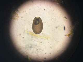
LIFE CYCLE [Duzynski, et al., 1998b]
Coccidia are present in the environment as oocysts, roughly
equivalent to the egg stage of other parasites. The oocysts are
usually passed from the host in the feces, and this is the form by
which almost all coccidia are known and identified. Eimeria
macusaniensis is distinct from the �small coccidia� by virtue of
its large size (~90 microns versus ~30 microns) and piriform
shape. Oocysts do not become infective unless environmental
conditions are appropriate, at which point sporocysts containing
sporozoites develop. The encapsulated oocyst is then referred to
as a sporulated oocyst, and it can persist in the environment in
this stage for years. The thick walls of sporulated E. mac make
this organism virtually impervious to environmental extremes and
it can persist in the soil for years. Some owners have resorted to
extreme means to attempt to destroy E. mac oocysts in their
fields, including topsoil removal and burning, but these measures
are neither effective [Cebra, 2007], nor likely to be warranted.
Once a sporulated oocyst is ingested by a host, the covering of
the oocyst is broken down either by mechanical or chemical
activity in the host�s gut. After release, the sporozoites seek
out and penetrate epithelial cells lining the gut. Inside the
host�s cells, the sporozoites begin to multiply asexually into
merozoites. The proliferation of merozoites eventually bursts the
host cell, and the released merozoites begin seeking out new cells
in which to begin the process of multiplication again.
Each species of coccidian is believed to have a specific number of
asexual replication cycles that it completes. The final generation
of sporozoites again seek out new host cells, but instead of
making more sporozoites by fission, they develop into two types of
gamonts: large, sessile macrogamonts (analogous to eggs) and
smaller microgametocytes, which produce motile microgametes
(analogous to sperm). When a macro- and microgamete fuse, a
protective wall develops around the resulting zygote, which pushes
free of the host cell and is excreted in the feces [Duzynski, et
al., 1998b].
The prepatent period, the time between ingestion of a sporulated
oocyst and passing oocysts in the feces, varies from species to
species of coccidia. The prepatent period for the two most common
small coccidia in alpacas are ten days for E. punoensis and 16-18
for E. alpacae [Foreyt, 1992]. Eimeria macusaniensis is unusual in
that its prepatent period is 33-42 days, meaning than an animal
can be infected for over a month before any trace of the parasite
is evident in the animal�s feces. This unfortunately means that an
animal may sustain considerable damage or even die from the
activity of the parasite before there is any means of detecting
the infestation.
ENVIRONMENTAL FACTORS PROMOTING OOCYST SURVIVAL
Oocysts are known to sporulate more quickly at higher temperatures
than lower, within the range of 50�F t o 122� F. Unsporulated
oocysts do not survive outside of these temperature extremes,
although sporulated oocysts can. Oocysts require moisture, oxygen
and shade to sporulate. Direct exposure to sunlight will kill
unsporulated oocysts [Duzynski, et al., 1998b]. Once sporulated,
oocysts remain infective for anywhere from several weeks to
several years in the natural environment, depending on species.
Eimeria macusaniensis requires 13-21 days to sporulate [Cebra, et
al., 2007].
In essence, coccidia thrive in damp, dark locations at moderate
temperatures � accumulated dung and bedding that do not dry out
are a haven for coccidia growth. You can limit your animals�
chances at (re)infection in several ways. Good manure management
is important: remove manure from animal living areas regularly and
consider allowing free range poultry to turn over your manure
piles, since exposing the oocysts to sunlight prior to sporulating
will kill them. The lengthy time period reqjuired for E. mac to
sporulate means that prompt manure removal can be very beneficial
in reducing the number of infective oocysts present in your
animals� environment.
The better overall health your animals are in, the better able
they are to resist infection and mitigate the effects of all
coccidia, including E. mac. Keeping stocking rates low and
removing stressors from your animals� environment will promote
good health and reduce the effects of coccidia. A good plane of
nutrition also permits an animal to carry a coccidia load without
significant ill effects. Over time, alpacas will build immunity to
the coccidia to which they have been repeatedly exposed, which is
why clinical cases of coccidiosis are usually seen in young
alpacas or older animals with compromised immune systems.
The same holds true for E. mac, although this species can be more
virulent than the small coccidia, particularly in cria. However,
it is entirely possible for an animal to carry subclinical loads
of Eimeria macusaniensis and remain in good health. Although E.
mac was initially viewed by many as a virtual death sentence, the
emerging consensus among camelid veterinarians is that E. mac
should be managed in the same way as small coccidia, but with
greater vigilance [Cebra, et al., 2007]. Cria are particularly
susceptible, and once an owner is aware of E. mac on their
property, the parasite must be considered in any animal showing
signs of ill thrift.
CLINICAL ILLNESS: COCCIDIOSIS
Coccidiosis caused by the �small coccidia� typically shows a
clumped or ball stool in mild cases, progressing to diarrhea and
weight loss in more severe infestations. In very severe
infestations, portions of the intestinal lining may be shed, and
the damage to the intestinal wall can be permanent, causing
continued ill thrift or stunted growth. Eimeria macusaniensis is
atypical in that diarrhea is not usually associated with even
heavy loads of the parasite. Weight loss and weakness are symptoms
of infection, but obviously are not diagnostic for E. mac alone.
Bloodwork from an infected animal will reveal hypoproteinemia (low
protein levels), but this is also not specific to E. mac
infection.
E. mac oocysts are large and heavy, and can be easily overlooked
in a fecal with a short float time, or one that is not
centrifuged. Eimeria macusaniensis also sheds very few eggs
(estimated at fewer than 100 in the first week of patency [Cebra,
2007]), so infrequent fecals are unlikely to find the parasite. If
E. mac is suspected, multiple fecal examinations over a several
week period, using centrifugation and a saturated sugar solution,
are recommended as the best way to try to catch oocysts as they
are shed.
TREATMENT
Animals with known or suspected E. mac can be treated in two
different ways: with a coccidiostat that prevents additional
reproduction of the coccidia, or with a coccidiocide, which kills
the organisms outright. Coccidiostats include amprolium (Corid),
which inhibits thiamine uptake in coccidia; sulfadimethoxine (Albon),
which prevents the uptake of folic acid; and sulfamethoxazole/trimethoprim
(SMZ-TMP), which also prevents the uptake of folic acid.
Without access to thiamine or folic acid, coccidia are unable to
continue reproduction, and the alpaca�s immune system will clear
the remaining organisms on its own. Although dosages and protocols
vary, most coccidiostats are used on an on-off-on rotation over
several weeks. It is important to note that alpacas are very
susceptible to thiamine depletion, much more so than other
ruminants. Thiamine depletion results in polioencephalomalacia (PEM),
characterized by swelling in the brain, which can be fatal.
Symptoms include lack of appetite, poor coordination and other
neurological signs. High doses of injected thiamine can reverse
PEM.
For this reason, if using amprolium to treat coccidiosis, it is
recommended that you concurrently administer thiamine
subcutaneously every third day during treatment. It may seem
counterintuitive to administer thiamine when amprolium works by
blocking access to thiamine (the coccidia preferentially uptake
the amprolium in place of thiamine). However, the injected
thiamine is available to the alpaca�s metabolism, but does not
reach the gut where the coccidia are.
Another important consideration when using coccidiostats in cases
of known or suspected Eimeria macusaniensis is that coccidiostats
are most effective against the first stages of a coccidial
infection. Given the long prepatent period of E. mac, these stages
may be past by the time treatment is begun. For this reason, a
coccidiocide may be preferable. Two coccidiocides are currently
recommended for the treatment of E. mac, ponazuril and toltrazuril.
Ponazuril (Marquis) is a medication originally developed to fight
a protozoan in horses. It is effective against later stages of
coccidial infection. The medication is very expensive and requires
careful dilution to an effective dosage for alpacas (40 gm
ponazuril paste plus 60 gm distilled water to equal 100 gm; dosed
at 9mg/lb once diluted). While equine veterinarians stock the drug
in many areas of the country, it may not be available except by
mail order in others. It must be administered for three days.
Toltrazuril (Baycox) is a relatively new treatment for
cocciodiosis. It is a coccidiocide, which kills the intracellular
life stages of coccidia. It must be imported from Australia, and
is available in this country from Light Livestock Equipment (www.lightlivestockequipment.com)
or can be ordered directly from Australian sources. Many farms
have reported success with single treatments, while others indicate that two doses several days apart are more
effective.
You should consult with your veterinarian to see which medication is
recommended for your particular situation. In mild cases of
clinical coccidiosis, coccidiostats may be preferred, as they do
permit the animal to mount its own immune response to the
nonreproductive coccidia. This will reduce the likelihood of
future reinfection. In severe cases, where immediate relief from
severe infection is required, a coccidiocide�s action may be more
appropriate. It is important that the alpaca community refrain
from overusing toltrazuril and ponazuril, in order to maintain
their effectiveness for the future.
Proper hygiene and good husbandry to prevent coccidiosis are
preferable to chemical intervention. Well cared for animals will
typically develop an immunity to coccidia � including Eimeria
macusaniensis. E. mac is now a widespread part of the parasite
community affecting North American camelids, just as are the small
coccidia. With proper management of your herd, the effects of this
parasite on your own herd, as well as that of others, can be
minimized. Proper quarantine and fecal testing of incoming and
outgoing animals should take into consideration the longer
prepatent period and low shedding rate of this organism, and
animals should remain in quarantine for an appropriate duration.
REFERENCES CITED
Cebra, C. K,. et al, �Eimeria macusaniensis infection in 15 llamas
and 34 alpacas,� J Am Vet Med Assoc, 230(1), 2007, pp. 94-100
Duzynski, D.W. et al., �The Coccidia of Camlidae,� 1998a, NSF-PEET
DEB 9521687, available as an on-line reference at http://biology.unm.edu/biology/coccidia/
artiodact2.html
Duzynski, D.W. et al., �Biology of the Eimeriidae,� available as
an on-line reference at http://biology.unm. edu/biology/coccidia/eimeriabiol.html
Guerrero, Carlos at al., �Eimeria macusaniensis n. sp. (Protozoa:Eimeriidae)
of the alpaca (Lama pacos),� Journal of Eukaryotic Microbiology,
18(1), 1971, pp. 162- 163
Jarvinen, J.A., �Prevalence of Eimeria macusaniensis (Apicomplexa:Eimeriidae)
in Midwestern Lama spp.,� Journal of Parasitology, 85(2), 1999,
pp. 373-376
Jarvinen, J.A., �Infection of Llamas with Stored Eimeria
macusaniensis Oocysts Obtained From Guanaco and Alpaca Feces,�
Journal of Parasitology, 94(4), 2008, pp. 969-972
Johnson, Amy et al., �Diagnosis and treatment of Eimeria
macusaniensis in an adult alpaca with signs of colic,� The
Veterinary Journal, 179(3), 2009, pp. 465-467
Palacios, C. A., et al., �Eimeria macusaniensis and Eimeria
ivitaensis co-infection in fatal cases of diarrhoea in young
alpacas (Lama pacos) in Peru,� Vet Rec., 158 (10), 2006, p. 344
Walker, Pam, �Gastrointestinal Parasites in Alpacas,� available
online at
http://www.alpacajack.com/ParasiteControl-for-your-Alpaca-Herd-81.htm
~~~~~~~~~~~~~~~~~~~~~~~~~~~~~~~~~~~~~~~~~~~~~~~~~~~~~~~~~~~~~~~~~~~~~~~~~~~~
From Parasite Management in Camelids by
Stacey Byers, DVM, MS, Dipl ACVIM
I. PROTOZOA A. Coccidia (Eimeria alpacae,
E. lamae, E. macusaniensis, E. punoensis)
This parasite typically causes diarrhea and weight loss or lack of
gain in crias and na�ve (previously unexposed) or immunosuppressed
adults. There is no cross protection between species so adults can
be infected and develop clinical disease from a different species.
Treatment is typically only recommended if oocyst (egg) counts are
significantly high with the presence of diarrhea. However if E.
macusaniensis is suspected, treatment is often started regardless
of finding oocysts in the feces since the parasite can cause
significant intestinal damage.
Infection occurs by oral exposure can occur in as little as 4 days
if oocysts are exposed to cool, moist conditions. Pasture
management is key to reduce exposure. The oocysts die in warm, dry
pasture in 20-30 days but can persist for years in cool, damp
environments. The prepatent period (time from ingestion of the
oocyst to shedding in feces) is variable among species but ranges
from 10 days for E. punoensis to over 40 days for E. macusaniensis.
Oocysts cause diarrhea by damaging intestinal cells. After the
anthelmintic treatment is finished, feces may remain loose until
the intestinal lining is repaired. In severe infections, stunting
or ill-thrift with continued diarrhea may occur due to permanent
damage to the intestinal lining.
|