|
Blood Transfusion in Llamas
from book: Veterinary Techniques in Llamas &
Alpaca by Dr. David Anderson
p. 333 - 335 by Meredyth L. Jones
Donated blood should not exceed 20% of the
blood volume of the donor which equates to about 1.5 of the donor's
body weight.
When the collection and administration volumes
are calculated, the donor is restrained in the standing position
(preferably in a chute) and the catheter connected to the
appropriate receptacle containing anticoagulants.
The container is lowered to the ground and
filled by gravity flow. In the case of commercial blood collection
bags, they should be filled until turgid and then rocked to assure
proper mixing of the blood and anticoagulant. Where these bags are
not available, any sterile receptacle may be used Sodium citrate is
added to the receptacle at a volume to create a 1:9 ratio of sodium
citrate : whole blood.
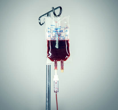
After the desired volume is collected, the bag
is attached to a filtered administration set (figure 73.2) and
administration to the donor initiated. The transfusion should begin
at a slow rate of 5 mL/kg/hour for the first 15-20 minutes and the
recipient monitored for signs of transfusion reaction. After this
time, if no abnormalities are noted, the rate may be increased to 10
mL/kg/hour for the remainder of the transfusion.
In cases where acute hemorrhage is the cause of
anemia, the bleeding must be stopped prior to or during the
transfusion because the volume expansion will worsen the losses. In
the case of hemolytic (relating to the disintegration of red
blood cells) disease, efforts should be made to identify the
cause and minimize hemolysis (the breaking down of red blood
cells with liberation of hemoglobin) because the average
lifespan of transfused red blood cells is limited to 3 to 5 days.
Severe anemia may be recognized during the examination by
inspection of the mucous membranes. Extreme anemia causes a pale
appearance. Assessment of the anemic patient should include
examination of peripheral blood. This may allow identification of
Mycoplasma haemalamae blood cells.
|