Foot and Mouth
Disease:
ARE LLAMAS AND ALPACAS AT RISK?
David E Anderson, DVM, MS, Diplomate ACVS
Associate Professor, Ohio State University Based on the inquiries I have gotten over
the past few weeks, I feel it would be timely to mention a few words
about the current scare in Europe with Foot and Mouth Disease (FMD). As
many of you know, I have been preaching biosecurity as an issue for the
future for the industry. You have only to talk to the llama and alpaca
owners in the United Kingdom to see how this can effect you whether you
like it or not!
Are llamas and alpacas at risk?
Unfortunately, the answer is both yes and no. Yes, llamas and alpacas
have been infected with FMD. No they do not appear to be very
susceptible to it. FMD infection in alpacas in Peru was confirmed in the
1970's. FMD risk in llamas and alpacas was researched in the USA and
Argentina. Routes of infection included tongue scarification,
intramuscular injection, intradermal injection, intravenous injection,
and cohabitation. Llamas and alpacas appear to be fairly resistant of
infection by natural exposure (cohabitation) but can and do succumb to
infection when any of the other exposure methods were used. Infected
llamas developed mild clinical signs including fever, anorexia, lesions
to the footpads, and lameness. Virus did not persist in any camelids
beyond 14 days after infection. Certainly, the risk of llamas or alpacas
becoming infected seems extremely low.
Fondevila et al studied the susceptibility
of llamas to FMD natural exposure in a biocontainment facility in
Argentina. This was a collaborative study between CICV and INTA in
Argentina and the USDA and APHIS in the United States. In that study,
llamas were exposed by cohabitation to FMD strains A-79, C-3, and O-1.
Of 30 llamas exposed to FMD virus infected pigs, only 3 showed any
evidence of infection and only 2 llamas (exposed to the O-1 strain)
showed any clinical signs of infection. No llamas exposed to A-79 or C-3
strains of FMD showed signs of infection. Clinical signs were extremely
mild. More importantly, FMD virus could not be recovered from any
specimen obtained from the infected llamas beyond 14 days post-exposure.
Ten cattle, 10 sheep, 10 goats, 10 pigs, and 30 other llamas were
exposed to the 30 llamas that had been exposed to the FMD virus infected
pigs. None of these animals showed any clinical signs of disease.
Lubroth et al studied the susceptibility of
llamas to FMD by cohabitation and by innoculation. This was a study
conducted by the USDA in abiocontainment facility. In this study, 3
swine, 1 bull, and 6 llamas were used. FMD virus strain A-24 was used.
In Group I, 1 llama was innoculated with FMD A-24 by giving 2 ml
intralingually, 2 ml intranasally, and 1 ml in the footpad. After 24
hours, 3 swine were introduced to the room for 7 days. All 3 swine
showed clinical signs of FMD after 3 to 4 days of exposure. The llama
showed fever, excessive salivation, lameness, and anorexia. In Group II,
one calf was innoculated with FMD A-24 by intralingual and intranasal
routes. After 24 hours, 2 llamas were introduced to the room. One of the
two llamas developed mild clincial signs of FMD as evidenced by fever on
day 2, and oral lesions noted on day 4. In Group III, one llama was
innoculated with FMD A-24 intranasally. After 24 hours, 2 llamas were
introduced to the room for 7 days. One of the 2 llamas developed mild
foot lesions only. FMD virus could not be detected in any llama beyond 8
days post-innoculation or post-exposure.
These two studies lead us to believe that
llamas are much more resistant to "natural" FMD virus
infection compared with cattle, sheep, goats, and pigs where the
morbidity of disease is expected to approach 100 %. Further and most
importantly, llamas do not appear to "carry" the virus for
prolonged periods of time as is seen with cattle, sheep, goats, and
pigs. Based on these studies, a policy of livestock separation (to
diminish high concentration cohabitation exposure risk) and quarantine
of all camelids with no movement or visitation would seem reasonable and
prudent.
What is it? FMD is a viral infection of
cloven-footed animals (virus is family Picronaviridae, genus Aphthovirus,
7 serotypes: A, O, C, SAT1, SAT2, SAT3, ASIA1, and at least 60 subtypes
- a very adaptable virus!). It most seriously effects cattle but swine,
sheep, and goats can be severely effected at times. The virus does not
appear to infect horses or people but there is a concern that any animal
may act to spread the infection. The plethora of serotypes and subtypes
makes effective vaccination extremely difficult because little
cross-protection exists between serotypes. This is one reason why
slaughter, where practical, has been used to control and eradicate the
disease. People do not appear to be susceptible to the disease unless
severely immunocompromised.
Where is it? FMD is enzootic to Africa,
Europe, Asia, Japan, Philippines, and South America. The spread of FMD
is a critical concern to countries that do not have it (e.g. North
America, Australia, and New Zealand). A good example of why FMD
vigilance is critical: FMD was introduced to Canada in the baggage of a
European immigrant. Britain suffered a massive outbreak in 1967-68
possibly as a result of feeding infected Argentine lamb to swine. That
outbreak was controlled and the disease eradicated as was a smaller
outbreak in 1980. The British survived that outbreak, you can be sure
they will survive this one! The last reported case in the USA was in
1929. Australia and New Zealand have never had a case of FMD. FMD was
eradicated from Mexico in 1954. Thus, all of North America is currently
free of FMD. Apparently the Darien Gap (between Columbia and Panama) and
prevented northern spread of diseased cattle from South America.
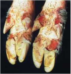
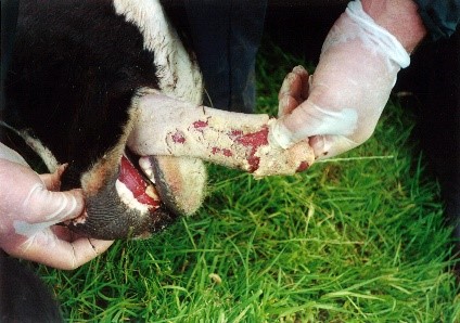
What does it do? FMD is most severe in cattle
and causes fever and vesicles in the mouth and on the feet. These cause
lameness and decreased feed intake because of pain. The virus takes from
1 to 7 days from the time of infection to the development of clinical
signs. At this time, high fever (104-106 F), low milk production, poor
appetite, and depression are noted. Excessive salivation is present and
vesicles (fluid filled pockets) are noted on the buccal mucosa, dental
pad, and tongue. The vesicles rupture within 24 hours leaving a painful
lesion. Vesicles also occur around the coronary band causing lameness.
As vesicles heal, animals return to eating over several days, but may
take up to 6 months to fully recover. Occasionally, the heart muscle is
damaged and acute deaths ensue. Diarrhea, sometimes including blood may
be seen. In sheep, goats and swine, the disease is usually much less
severe.
How deadly is it? FMD rapidly spreads within a
herd and essentially 100% of susceptible animals succumb to the disease.
FMD is not considered a particularly lethal disease. Death rates rarely
exceed 2% in adults and 20% in young stock. There have been outbreaks
with up to 50% mortality. However, prolonged convalescence causes severe
losses in production and health, cripples animal industries, and
severely inhibits travel and tourism. Where does it come from?
There are a variety of species that allow the virus to persist or serve
to spread the infection. Some include elephants, capybara, hedgehogs,
coypu, rodents, birds, and wild ruminants (Roe deer, muntjac, sika deer,
fallow and red deer, water buffalo). These animals may not show clinical
signs, but may harbor the virus to allow later spread of the infection
to susceptible species. These species are not likely to play a major
role in transmission because of lack of contact with susceptible
species. Sheep may carry the virus for up to 5 months. African buffalo
may harbor the virus for up to 28 months! Goats may also serve as
carriers of the disease. One study in Kenya showed that goats served a
minor role in transmission to cattle and that sheep were not significant
carriers. In other outbreaks, sheep meat imported from infected areas
appear to have been the origin of infection.
How is it spread? The virus may be spread by
inhalation or ingestion. Initial outbreaks are most commonly caused by
ingestion (e.g. infected meat), but rapid spread within a herd is likely
via inhalation (airborne virus). Wind and humidity appear to increased
windborne spread. Virus spread has been estimated to be as far as 62
miles (100 kilometers)! Up to 50% of infected animals may remain as
carriers of the disease for at least 6 months. Virus could be recovered
from nasal secretion of PEOPLE for up to 28 hours after working with
infected cattle. In England, one estimate of how the disease was spread
included birds (16%), meat products in pig food (40%), meat and bones
(7%), unknown (7%), and obscure (28%).
Can we kill the virus? FMD is a very stable
virus. It can survive up to 1 year in the environment, 10 to 12 weeks on
clothing and feed, and 30 days on hair! Sunlight, boiling, and
autoclaving rapidly destroy the virus. Most disinfectants and meat
packing industry techniques do not destroy the virus. If you travel in
an area that has FMD, you should use disposable shoes and clothing (e.g.
coveralls), shower extensively after the visit and before traveling, and
preferably stay away from any farm for at least 30 days. The best bet is
to stay clear of infected areas during active outbreaks of disease.
Do animals become immune? Cattle mount an
effective immune response to FMD that lasts up to 4 years. Swine
immunity persists for only 7 to 8 months. Immunity is relatively
specific to the serotype involved in the exposure. New outbreaks with
different serotypes can occur at any time. How is it diagnosed?
There are multiple tests that have been used including tissue culture,
virus neutralization, compliment fixation tests, experimental infection,
and ELISA tests. A government-approved laboratory must perform these.
FMD is a federally reportable disease in the USA.
Is there a vaccine? Yes, but success of
vaccination programs has been highly variable because of the multitude
of serotypes and subtypes. The most common types are killed virus
trivalent forms. Vaccination in the USA is not permitted. Suspected
cases of FMD are required to be reported to federal authorities for
investigation and immediate responses to control spread.
Reading:
Fowler ME. Medicine and Surgery of South American Camelids. Iowa
State University Press.
Blood DC, Radostits OM. Veterinary Medicine (7th Edition).
Bailliere and Tindall.
Lubroth J, Yedloutschnig RJ, Culhane VK, Mikiciuk PE.
Foot-and-mouth disease virus in the llama (Lama glama): diagnosis,
transmission, and susceptibility. J Vet Diagn Invest 1990;2:197-203.
Fondevila NA, Marcoveccio FJ, Viera JB, O'Donnell VK, Carrillo BJ,
Schudel AA, David M, Torres A, Mebus CA. Susceptibility of Llamas (Lama
glama) to infection with foot-and-mouth disease virus. J Vet Med
1995;B42:595-599.
David E Anderson, DVM, MS
Diplomate, American College of Veterinary Surgeons
Associate Professor of Surgery, Food Animal
601 Vernon L Tharp Street
College of Veterinary Medicine
The Ohio State University
Columbus, Ohio 43210
|